Radiologists Hope to Use AI to Improve Readings
The University of Miami Miller School of Medicine Department of Radiology is working with the University’s Institute for Data Science and Computing (IDSC) to design an artificial intelligence tool that could help them diagnose patients in a more individualized way. Over the years, new technology has helped radiologists diagnose illnesses on a multitude of medical images, but it has also changed their jobs.

While in the past these physicians spent more time speaking with patients, today they spend most of the time in the reading room — a dark space where they scrutinize images alongside a patient’s electronic medical records and other data sources — to diagnose an illness.
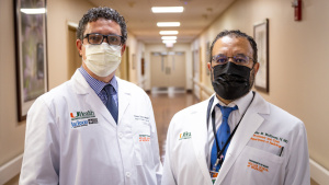
A radiologist’s job is often solitary. And it is a trend that Miller School radiologists Alex McKinney, M.D., chair of the Department of Radiology, and Fernando Collado-Mesa, M.D., hope to change.
The two physicians have been working with the IDSC to create an artificial intelligence toolbox that will draw on a massive database of deidentified data and medical images, to help doctors diagnose and treat diseases based not only on imaging data but also by considering a patient’s unique background and circumstances. This would include risk factors such as race and ethnicity, socioeconomic and educational status, and exposure. The physicians say it is a necessary innovation at a time when narrow artificial intelligence in radiology is only able to make a binary decision such as positive or negative for one disease, rather than scanning for a host of disorders.
“We believe the next iteration of artificial intelligence should be contextual in nature, which will take in all of a patient’s risk factors, lab data, past medical data, and will help us follow the patient,” Dr. McKinney said. “It will become a form of augmented interpretation to help us take care of the patient.”
Putting Data in Context of the Individual Patient
According to Dr. Collado-Mesa, “This toolbox will not just say yes or no, disease or no disease. It will point to the data around it to consider a variety of issues for each individual patient, to put its findings into a context, including future risk.”
Current artificial intelligence tools are also limited to a specific type of medical image, and cannot, for example, analyze both MRI (magnetic resonance imaging) and ultrasound at the same time. In addition, the patient data that is used in these diagnosis tools is typically not inclusive of a range of demographic groups, which can lead to a bias in care. Having a tool that draws upon the examples of millions of South Florida patients while maintaining their privacy will help radiologists be more efficient and comprehensive, Dr. McKinney noted.
“Right now, there is just so much data for radiologists to sift through. This could help us as our tech-based partner,” Dr. McKinney added.
A Better Alternative
All of these factors led Drs. Collado-Mesa and McKinney to try to create a better alternative, and they spoke with Nick Tsinoremas, Ph.D., IDSC IDSC director, and professor of biochemistry and molecular biology. Dr. Tsinoremas and IDSC’s advanced computing team came up with the idea of utilizing an existing tool called URIDE — a web-based platform that aggregates deidentified patient information for faculty research — and adding in the deidentified images from the Department of Radiology.
They hope to unveil a first version of the toolbox this summer, and plan to add new elements as more imaging data is added. It will include millions of CT scans, mammograms, and ultrasound and MRI images, along with radiographs, Dr. McKinney pointed out.
“We don’t want to rush this because we want it to be a high-quality, robust toolbox,” said Dr. Collado-Mesa, an associate professor of radiology and breast imaging, as well as chief of innovation and artificial intelligence for the Department of Radiology.
Both physicians and Dr. Tsinoremas hope that the artificial intelligence tool will help answer vital research questions such as: “What risk factors lead to certain brain tumors?” and “What are the most effective treatments for breast cancer in certain demographic groups?” It will also use machine learning, a technique that constantly trains computer programs how to utilize a growing database, so it can “learn” the best ways to diagnose certain conditions.
“Creating this resource can help with diagnosis and will allow predictive modeling for certain illnesses, so that if a person has certain image characteristics and clinical information that is similar to other patients from this database, doctors could predict the progression of a disease, the efficacy of their medication, and so on,” Dr. Tsinoremas said.
Adding to Data and Scope
To ensure the toolbox will be unbiased, the team is also planning to add more images and data of all population groups in the community, as it is available, as well as to monitor the different elements constantly and systematically within the toolbox to make sure it is performing properly.
The radiologists plan to focus first on illnesses that have a high mortality or prevalence in the local population, like breast cancer, lung cancer, and prostate cancer, and to add others with time.
The technology could allow them to spend more time with patients and offer more personalized, precision-based care based on the patient’s genetics, age, and risk factors, according to both physicians.
“Artificial Intelligence has the potential to advocate for the patients, rather than a one-size-fits-all approach to medicine based on screening guidelines,” Dr. McKinney said. “This could help us get away from that, and it would hopefully offer more hope for people with rare diseases.”
But as data is added in the future, the researchers hope to expand their work with the tool. And they hope that physicians across the University will use it to conduct medical research, too.
“This is a resource that any UM investigator could potentially access, provided that they have the approvals, and it could spark a number of different research inquiries to describe the progression of disease and how patients respond to different treatments in a given time period — these are just some of the questions we can ask,” Dr. Tsinoremas said.
Tags: artificial intelligence, Department of Radiology, Dr. Alex McKinney, Dr. Fernando Collado-Mesa, Dr. Nick Tsinoremas, University of Miami Institute for Data Science and Computing